Ventricular Septal Defect
What is a ventricular septal defect?
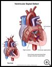
A ventricular septal defect (VSD) is a hole in the heart that forms an abnormal connection between 2 of the heart's chambers.
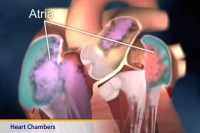
There are 4 chambers in the human heart. The 2 lower chambers are called the right and left ventricles, and the upper 2 are called the right and left atria. The wall between the 2 ventricles is called the ventricular septum.
When there is a hole in the ventricular septum, oxygen-rich blood from the left ventricle flows into the right ventricle. Then it is pumped back to the lungs, even though this blood does not need oxygen. This problem makes the heart have to work harder to pump blood.
How does it occur?
A VSD is found most often in babies. It happens before birth and is the most common birth defect of the heart. Often it is the only defect, but sometimes there are other heart defects as well. Most of the time the cause of the birth defect is not known. A gene defect or something else may keep this part of the heart from developing properly.
Rarely, an injury to the heart may cause a VSD.
What are the symptoms?
Septal defects vary in size and in the symptoms they cause. A small VSD usually causes no symptoms. Even if the defect is large, a baby often does not have symptoms until several weeks after birth. Some babies with a large VSD do not grow normally and may become undernourished because they do not feed well. Other symptoms include sweating, increased breathing rate, and frequent lung infections. A large VSD in small children can lead to severe heart failure. This means that the heart cannot do its proper job as a pump.
How is it diagnosed?
Your healthcare provider will listen for a heart murmur with a stethoscope. A heart murmur is an extra sound heard between heartbeats. The murmur is caused by the blood flowing abnormally through the heart. Sometimes, if the opening is very large, a murmur may not be heard.
Your child will have a physical exam, and the following tests may be done:
- a chest X-ray to look for enlargement of the heart
- an electrocardiogram (EKG), which is a recording of the electrical activity of the heart
- an echocardiogram, which is a type of ultrasound scan
- heart catheterization, which is a procedure for passing a very thin tube through a blood vessel into the heart
In addition to making pictures of the heart, an echocardiogram can show if there is increased blood pressure in the lungs. Catheterization may be used to confirm the diagnosis and to make sure there are no other heart problems. It can also be used to repair the heart.
How is it treated?
A small VSD usually does not cause any problems and seldom needs treatment. Small VSDs may close on their own during the first years of childhood. The smaller the defect, the greater the chance that it will close on its own. People with a small VSD may lead normal lives.
Medium and large ventricular septal defects may need to be fixed with surgery or heart catheterization. The VSD is closed by sewing a patch of a special material (Dacron) over the defect. This treatment helps prevent problems later in life, including heart failure and high blood pressure in the lung arteries.
If your baby has symptoms, medicines will be given to help the heart pump better until your child has surgery.
When surgical repair of a VSD is not an emergency, the operation carries very little risk.
How long will the effects last?
Often, if the VSD is small, it will close on its own. If a large VSD is not repaired, a child may have trouble growing. The child may have signs and symptoms of heart failure--for example, breathing problems.
Children whose VSD is repaired before they are 2 years old usually do well. Older children and young adults may still have some problems with how their heart works after the defect is repaired. These problems, which include abnormal heart rhythms and a slightly reduced pumping ability of the heart, are usually not serious and may be treated with medicine.
Last modified: 2011-08-08
Last reviewed: 2011-06-06


