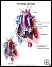
Tetralogy of Fallot
What is tetralogy of Fallot?
Tetralogy of Fallot is a birth defect. This defect consists of 4 problems with the heart:
- a hole between the 2 lower heart chambers (the ventricles)
- an aorta that sits just over the hole between the lower heart chambers. The aorta is the artery that normally carries oxygen-rich blood from the left ventricle to the rest of the body.
- an enlarged right ventricle.
- narrowing under or at the pulmonary valve. This valve is the opening between the right ventricle and the artery that carries blood to the lungs. This defect can also involve the lung arteries.
The hole between the ventricles means that oxygen-rich blood mixes with oxygen-poor blood. Narrowed pulmonary arteries and pulmonary valve means less blood can reach the lungs. These defects make the right ventricle work extra hard. It gets bigger and thicker. When oxygen-poor blood flows through the aorta to the body, your child’s skin looks blue.
How does it occur?
Babies with this condition are born with it. What causes it is not known.
What are the symptoms?
Children with this birth defect often look blue. The blueness may appear at birth or soon after. Babies may have spells during which they turn blue, breathe rapidly, and cry. They may pass out. During exercise, older children who have not had surgery often get short of breath and may tire easily. They may squat during exercise to help them catch their breath. These symptoms happen because the children are not getting enough oxygen. Not enough blood is flowing to the lungs to pick up the oxygen their body needs
How is it diagnosed?
Children with this defect usually have a heart murmur, which a healthcare provider can hear with a stethoscope. A heart murmur is an extra sound made between heartbeats. These murmurs are caused by the blood flowing abnormally through the heart.
To diagnose the problem, the following tests may be done:
- an echocardiogram, which uses sound waves to make pictures of the heart
- heart catheterization, which is a procedure for passing a small tube through an artery of the leg and into the heart to take special X-ray pictures of the heart with a dye.
How is it treated?
To correct the problem, surgery is done to close the hole between the two lower heart chambers. This usually requires a patch of synthetic material such as Dacron.
The surgeon will also relieve the narrowing at the pulmonary valve. This may be done in several ways:
- The pulmonary valve may be replaced with a new artificial or tissue valve.
- The surgeon may enlarge the area of narrowing of the heart with a patch of synthetic material or a thin layer of tissue from the sac surrounding the heart (the pericardium).
- If a baby is too sick or the pulmonary arteries are too small, a small tube is placed between one of the branches of the aorta and the pulmonary artery. This tube is called a shunt. It lets blood enter the lungs and allows the lung arteries to grow. Corrective surgery can be done at a later time.
How long will the effects last?
After surgery, most children are able to do all normal things, including sports. Some children need to limit their activities. Some may need to take medicine to control their heart rate and to help their heart pump better. Later in life, they may have abnormal heart rhythms.
Children with tetralogy of Fallot should see a cardiologist who specializes in congenital heart disease regularly for the rest of their lives.
Ask your healthcare provider if your child should take antibiotics to prevent infection before having dental work or procedures that involve the rectum, bladder, or vagina.
Last modified: 2011-05-26
Last reviewed: 2011-05-19

